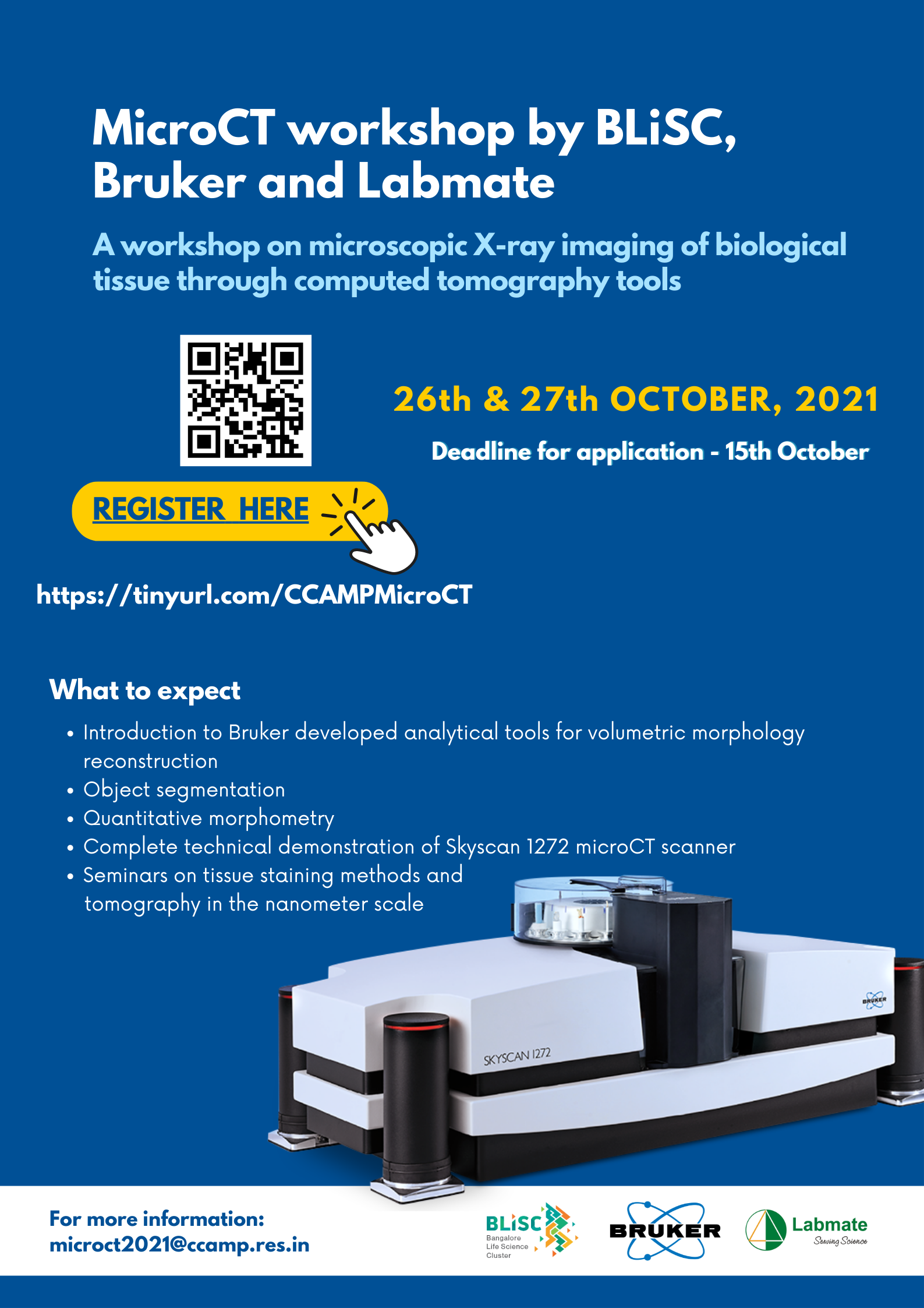
Dates: Tuesday, 26th - Wednesday, 27th October, 2021
Times: Half a day on both days [See Agenda below]
About the Course: This online workshop will introduce you to microscopic X-ray imaging of biological tissues through computed tomography techniques. It will comprise of a series of webinars to discuss Bruker developed analytical tools for volumetric morphology reconstruction, object segmentation and quantitative morphometry. This includes a complete technical demonstration of biomaterial imaging and testing features in the latest Bruker Skyscan 1272 microCT scanner. Emerging advances in the resolution of X-ray volumetric imaging will be discussed through seminars on tissue staining methods and tomography in the nanometer scale.
Eligibility: Anyone from graduates to PhD students, postdocs, faculty, doctors and industry can apply. Seats not limited.
A course fee of 1000 ₹ incl. GST associated with the program. Please note that your registration will be confirmed only upon receipt of full Fee. Kindly arrange for Online Transfer to our bank account. (Details of online transfer in registration link). Please send transaction details for the payment like, date, amount and transaction ID once payment is processed.
Register: https://tinyurl.com/CCAMPMicroCT by 15th October, 2021
Feel free to get in touch with us over e-mail at microct2021[at]ccamp.res.in for any further queries.
Agenda -
Speakers:
 Kjell Laperre, PhD- Kjell Laperre is the Market Product and Applications Manager, responsible for life science microCT products at Bruker Preclinical Imaging (PCI). Kjell holds a Master’s degree in Biomedical Sciences and obtained a PhD degree in the department of Experimental Medicine and Endocrinology at the Catholic University of Leuven in Belgium. In 2012, Kjell joined Bruker as life science application scientist. Bruker Belgium, formerly known as SkyScan, develops and produces a wide range of high-end microtomography instruments for life science, material research and in vivo preclinical studies. Since 2015, Kjell took the position of applications manager in Bruker microCT and further evolved into Market Product Management in 2018.
 Philip L. Salmon, M.Sc., Ph.D - Dr Phil Salmon is currently a Biomedical Application Scientist at the micro-CT manufacturer Bruker-microCT Belgium, responsible for the development and validation of systems and software for micro-CT morphometry, densitometry, model visualization and other biomedical applications. He received two M.Sc. degrees from Plymouth Polytechnic and St Andrews University, Scotland, UK, and his Ph.D. at Bristol University, UK. In his current role at Bruker-microCT for two decades, he has been directly involved in the practical implementation of the new technology of microCT imaging and 3D analysis in bone and dental biology, as well as in other life science and general research fields. He has played a leading role in the development of the “CT-Analyser” software which is a global standard for 3D quantitative morphometry and micro-densitometry. He invented the “post-scan” method for movement correction in micro-tomography which is being applied world-wide in micro-tomographic and synchrotron imaging. He remains active in research and has authored a comprehensive study of the beam hardening-associated context sensitivity of microCT densitometry, as well as host editing a research topic for the journal “Frontiers in Endocrinology / Bone Research” on pattern formation in bone. He makes frequent scientific presentations at international conferences and has published numerous peer-reviewed scientific papers.
 Ali Bahadur - Ali Bahadur is an Applications Scientist with the Preclinical Imaging division of Bruker Biospin Corporation which offers a wide range of modalities including MRI, MicroCT, PET, SPECT, and Optical Imaging. Ali has worked in the field of Preclinical Imaging for more than a decade encompassing both academia and industry, mainly focusing on the application and use of preclinical imaging in the drug discovery and development process. He has published many peer-reviewed papers in international scientific journals and has several granted patents that cover his original work in imaging technologies. At present, Ali is based at Bruker’s headquarters in Billerica, MA and provides product training and applications support to its Preclinical Imaging users worldwide.
 Jolien Dellafaille - Jolien Dellafaille is a microCT Application Scientist for the life sciences working within the Preclinical Imaging division (PCI) of Bruker. She completed her master’s as Veterinary Surgeon with focus on research and medical imaging at the University of Gent in 2018, she started working at Bruker Belgium in the beginning of 2019. Bruker Belgium develops and produces a wide range of qualitative microtomography instruments who can be used for non-destructive, high resolution 3D and 4D imaging. It is at this office and production site in Kontich, Belgium that she is located. She focusses on training and application support to all who work with the Bruker microCT systems.
 Prof. Dr. Javier Alba-Tercedor Prof. Dr. Javier Alba-Tercedor is a full Professor of Zoology at the Department of Zoology of the University of Granada in Spain, where he has been teaching different subjects (General Zoology, Entomology, Invertebrates, and Conservational Biology and Ecology of Water Courses) since October 1st of 1977. He conducted research and did stays in institutions and universities of different countries: Poland, Check Republic, Australia, Canada, and USA. The main topics of his research have been on aquatic macroinvertebrates: taxonomical and ecological aspects (mainly with insects, and mostly on Ephemeroptera and Plecoptera), describing new species from Europe, North and South America, Africa, and Australia. A lot of effort of his carrier it has been the use of aquatic macroinvertebrates for bioassessment of watercourses degradation. Thus, he developed the method IBMWP (Iberian Biomonitoring Working Party) nowadays official to assess the Ecological Status of the Spanish water courses to achieve the exigencies of the European Frame Water Directive, and he participated as scientific expert during the preliminary development that directive. As a result, he has published more than 250 titles (papers, books, chapters, reports, press news, etc.) on these subjects. However, he recognizes that since he first glimpsed the possibilities of micro-CT, and since in 2010 he started to work with microtomography, micro-CT has become his main passion and he has been doing research with this technique non only for anatomical studies, but what he likes to says as “unveiling mysteries in science”. Thank to these micro-CT works he was awarded several times during the annual international Skyscan/Bruker micro-CT user’s meetings he participated (best micro-CT picture, video, or best presentation). But he stresses that without a doubt the most curious experience that his passion for microtomography has led to him it was the collaboration with the film industry. Thus, he collaborated with the Canadian BUF’s visual effects company by scanning the beetles appearing in the film “Blade Runner 2049” that won 2018 Best Visual Effects Oscar in Hollywood. Most of his recent micro-CT papers have been publish in the journals PLoS One and Scientific Reports of Nature and many of his micro-CT videos can be visualized in his YouTube channel: http://youtube.com/albatercedor
Talk Title: NEW REVIVAL OF ENTOMOLOGICAL STUDIES: THE MICRO-CT AS A TOOL STRADDLING SCIENTIST RESEARCH, ART AND EDUCATION
High resolution microtomography (micro-CT) despite it is not a new technique, nowadays it is being used routinely in science. Mostly it is being used as a substitute to microscopy to look at/for external and internal structures. However it can be used to clarify non-answered questions, and also for educational purposes. And in many cases simply it represents itself, Art. The main advantage is that it is a non-destructive technique, and once the sample has been scanned, the reconstructed images can be stored, and at any time viewed, rotated, sliced, etc…, making it possible to study details of external and internal structures in any possible perspective. Here it will be explained what it is the microtomography, and how it works, presenting examples and giving details of how the author uses a high resolution Skyscan 1172 micro-CT for anatomical studies, and to unveil “mysteries” in Entomology . The talk will be illustrated with high resolution images, and videos to demonstrate how thanks to micro-CT it is possible to do amazing trips inside the insects. Moreover it will explained how the obtained “real” entomological models can be visualized, rotated, cut, etc…, in an interactive mode, when using mobile devices (smartphones and tablets), resulting very attractive for the students, and thereafter very promising for educational purposes. In any case, many of the obtained results have a nice visual artistic appearance, and attenders will understand why the Micro-CT represents a tool straddling scientist research, art and education.
 Dr. Naibedya Chattopadhyay - Naibedya Chattopadhyay, Ph.D. in Endocrinology from Sanjay Gandhi Post-graduate Institute of Medical Sciences in 1994, did his postdoctoral fellowship from Harvard University, where he later became Assistant Professor. He returned to India in 2006 and is presently the Chief Scientist, Division of Endocrinology at CSIR-Central Drug Research Institute and also a Professor of Biological Sciences, Academy of Scientific and Innovative Research (AcSIR). He is also the Director of Center of Excellence in Research in Anabolic Skeletal Targets in Health and Illness (ASTHI, CSIR). His research interests include bone biology, metabolic bone diseases and endocrine-related cancers. Naibedya is an Editorial Advisory Board Member of Biochemical Pharmacology (Elsevier) and American Journal of Physiology since 2008, Guest Editor of Special Issue of Current Molecular Pharmacology (Theme: Molecular and Pharmacological Aspects of Existing and Experimental Bone Anabolic Therapies, published in June 2012), and an Editor of a Book - Calcium-sensing Receptor; Kluwer Academic Publishers; Boston, MA, USA (2003). He has 208 publications with number of citation >11,500 and h-index of 63. He has eight patents and out-licensed three technologies to the industry out of which one is commercialized.
Irrfan Qayoom - Mr. Irfan Qayoom is a PhD student at Department of Biological Sciences and Bioengineering, Indian Institute of Technology Kanpur working under the supervision of Professor Ashok Kumar. He has done his Masters in Biotechnology from University of Kashmir, Srinagar, Jammu and Kashmir. His thesis is mostly directed towards the development of nanohydroxyapatite based ceramic implants as a drug delivery vehicle for the treatment of bone infections. He has been working using microCT (SkyScan 1172, Bruker) system for characterisation of scaffolds and evaluation of bone mineralization in rat models.
Dr. Dhananjay Chaturvedi - Dhananjay is a developmental biologist by training, having investigated molecular aspects of murine eye development and chromatin modifiers of Germline stem cells in Drosophila at TIFR Mumbai and UT Southwestern Dallas. In his post doc at NCBS he co-reported adult muscle stem cells in Drosophila for the first time. Ever since, he has employed CT scanning to see the effect genetic and physical manipulation of muscles in fruit flies. Most recently he employed CT scanning to elicit a longitudinal profile of Anopheles mosquitoes. He expects to start his own lab addressing interesting questions in Biology very soon.