
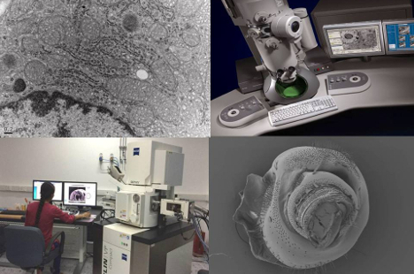
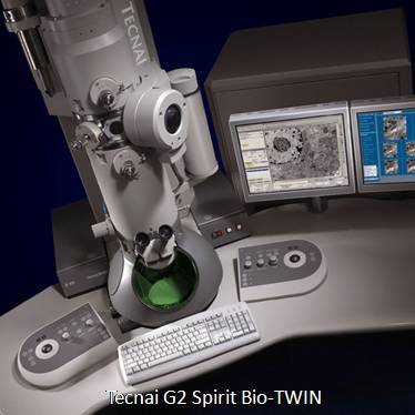
 Transmission Electron Microscope or TEM (Tecnai G2 Spirit Bio-TWIN )
- High contrast, high resolution for 20 –120 kV operation Accelerating Voltage 20, 40, 60 80 100 120 kV
- Beam source: LaB6 filament.
- Vacuum system: Oil Diffusion (ODP) and Rotary for projection chamber; Ion (IGP) and Turbo vacuum pumps for column.
- +/-80˚ fully computer controlled Goniometer stage
- Optimized for 2D & 3D imaging of cells, cell organelles and soft matter
- Smart Tracking Position System for sample navigation Low dose mode available
- Cryo attachment for imaging of biological specimens and materials samples at sub-zero temperatures
- 4k x 2k Gatan Orius side mount CCD camera.
- DM3 operation software (Gatan Digital Micrograph)
- 3D reconstruction software
- Holders: Standard single tilt; Gatan multiple specimen holder; Standard single tilt +/- 80˚ Tomography specimen holder; Gatan cryo holder single tilt +/- 80˚ Tomography Cryotransfer Holder
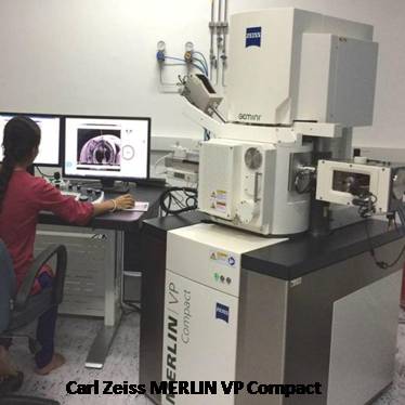 Scanning Electron Microscope or SEM (The MERLIN Compact VP)
- Resolution: 0.8nm @ 15KV; 1.4nm @ 1 KV; 0.8nm @ 30KV (STEM mode)
- Probe current: 5 pA to 100nA
- Acceleration voltage: 0.02 V to 30 kV
- Magnification: 12 – 2,00,000 X
- Electron Emitter:Thermal (Schottky) field emission gun.
- Detection Modes
- Correlative Microscopy with Shuttle and Find.
- Cryo SEM and freeze fracture for imaging hydrated samples or for beam/vacuum sensitive specimens.
- EDAX for chemical composition analysis and element mapping.
- STEM: Low voltage optimized bright field, 4 quadrant dark field, and high angular dark field transmission imaging.
- Variable Pressure: High efficiency VPSE detector for pressure up to 60 Pa
- Secondary Ion: High efficiency in-lens SE detector Everhart Thornley Secondary Electron detector
- In-lens Duo: Switchable between on-axis in-lens secondary electron detection for highest surface sensitivity and energy selective backscattered detection for advanced materials contrasts.
- Specimen Stage: 5-Axes Motorized Eucentric Specimen Stage:X = 130mm; Y = 130mm; Z = 50mm; T = - 3º to 70º; R = 360º (continous)
- Chamber: 330mm(Ø) x 270mm(h) 15 accessory ports for various options including CCD-Camera with IR-illumination
- Vacuum system: High vacuum mode and variable pressure mode
High-resolution 3D X-Ray Scanner or MicroCT (Bruker SkyScan 1272)